A New Way to Treat Strokes
OMS II Maxwell Horowitz Delivers Poster Presentation at Stroke Conference

A TouroCOM Harlem student presented a groundbreaking new approach to treating strokes at this year’s International Stroke Conference.
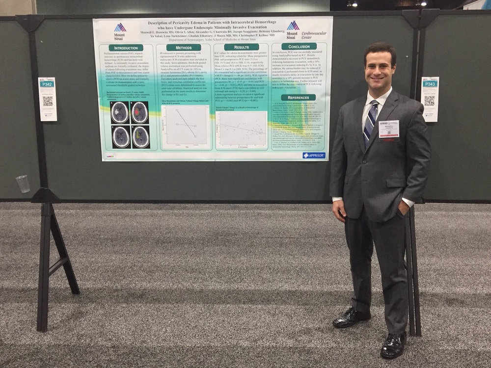
OMS II Maxwell Horowitz spent part of last summer working in neurosurgery department of the Icahn School of Medicine at Mount Sinai. Together with several leading neurosurgeons, Horowitz examined 48 intracerebral hemorrhage stroke patients who underwent endoscopic minimally invasive evacuation. He presented an abstract on the findings at the International Stroke Conference, held in California in January.
An intracerebral hemorrhage is caused by the burst of unhealthy blood vessel in the brain. The blood then leaks into the surrounding area and can become a hematoma, a solid swelling of clotted blood. Perihematomal edema is fluid caused by the disruption of the blood vessels and the toxic effects of the blood and clotting factors. Intracerebral hemorrhage affects almost 200,000 people in the US. Complications ensue afterwards: 25 percent of patients die, and 50 percent are functionally dependent afterwards due to severe neurological deficits.
In most cases after an intracerebral hemorrhage, doctors allow the stroke to heal itself by reducing blood pressure through medication or intracranial means or performing a craniotomy, removing a portion of the person’s skull to ease the pressure and allow access to the hematoma. However, in the technique that Horowitz studied, a doctor drills a small hole in the patient’s skull and then sucks out the hematoma. (Since the hematoma is sucked out, the edema is now called pericavity edema, meaning that the edema, the fluid, surrounds what is now a cavity in the brain.)
“Essentially, it’s like sucking up the blood with a straw,” Horowitz said. The results have been promising, according to Horowitz, with possible shorter recovery times and doing away with the need to remove a portion of the skull. The results indicated that patients had an immediate reduction in pericavity edema following treatment and had less edema growth in the first 48 hours compared to patients in comparable nonsurgical studies.
“We’ve been looking at whether the surgery reduces the edema burden,” Horowitz explained. “We use a specific program to look at the CT imaging and we’re able to use the program to measure, based on the contrast, what’s blood and what’s edema, what is the cavity and what is the air.”
Through the use of CT scans, the research team also discovered that the faster the surgery was performed after the incident of the stroke, the less fluid buildup left in the skull afterwards. (Preliminary results indicate that a day’s delay added a 10 percent increase to edema build-up).
The research team was flown out to California to the conference. Maxwell said he enjoyed the event. “I saw a lot of cool things and heard a number of great talks,” he said. Another highlight was when the doctors played Game of Strokes, a quiz game with conference attendees buzzing in answers to stroke-related questions.
“I tried my best but I’m not a neurosurgeon yet,” laughed Maxwell.
Yet, of course, being the operative word.

