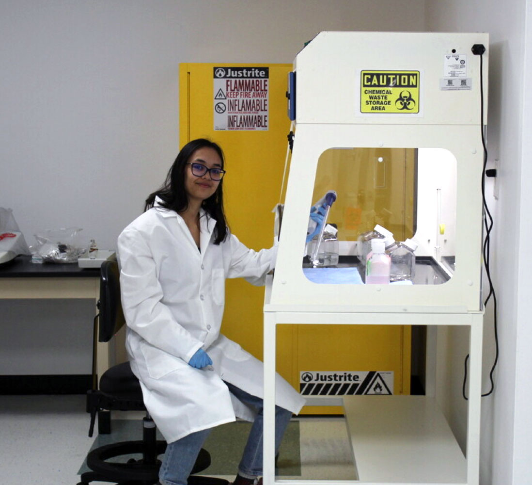
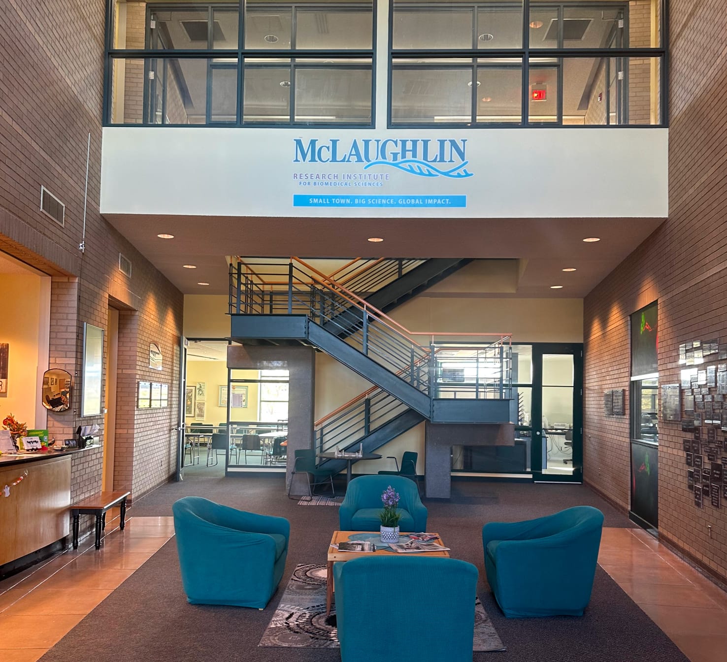
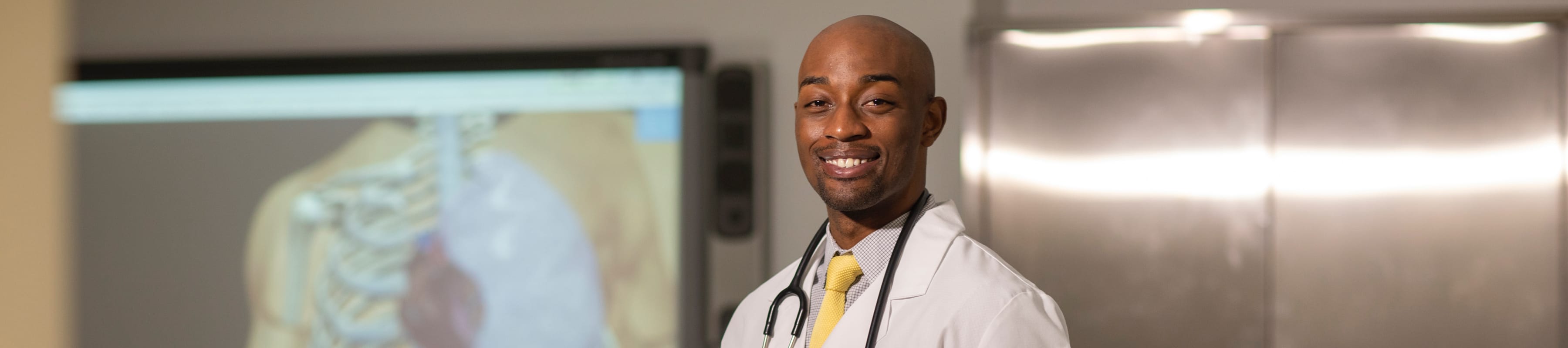
Research

To combat and prevent disease, we must first understand it. With fully equipped labs at our disposal, TouroCOM faculty are hard at work on innovative medical and educational research.

TouroCOM faculty looking for help in pursuing research projects can review available research resources. We're committed to continued research with a strategic plan for increased research at TouroCOM. 

Research Laboratories

TouroCOM facilities currently include labs that support advanced research in:

Cancer Biology Research Laboratory

This laboratory is equipped with variety of laboratory equipment including centrifuges, refrigerators, freezers, electrophoresis equipment, water baths, tissue culture incubators, biohazard safety cabinets, microscopes, etc., with bench space for six people. The lab is also equipped for primary epithelial cell culture, tumor biology, animal work, biochemistry, and cell biology. Equipment includes a Bio Rad CFX96 real time PCR machine, Bio Rad T1000 thermocycler, Amaxa transfection system, Eppendorf Biospectromoter, and Gene Pulser X-cell electroporator. There is access to communal equipment rooms, tissue culture rooms. The lab contains 110V and 220V emergency outlets with backup power as well as an emergency shower and eyes washes. Adjacent to Cancer Biology lab is the Changing/Autoclave Utility Room (Room 508) and bathroom.

Vivarium

These two animal holding rooms, one for mice and one for rats, are equipped with two Thoren systems with 50 Individually ventilated Microisolation cages for each system, 4 Baker biohazard safety cabinets, and a pass through autoclave for cage sterilization is available in room 508.

Research BioBank

This laboratory was established in 2011 in order to provide samples of fixed and frozen human tissues and fluids obtained and stored in a safe and ethical way. Those samples are acquired, processed and distributed to TouroCOM researchers for research projects on variety of human diseases and conditions where the human tissue is the sole source of information. This laboratory is equipped with -80deg C ultra-freezers, liquid nitrogen freezers, biosafety hoods, microscopes and a variety of small laboratory equipment.

Tissue Culture Laboratory

This laboratory is equipped with a full complement of equipment to establish, maintain and propagate cell and tissue cultures. It is equipped with Hepa-filtered bio-safety cabinets, CO2 incubators, hoods, microscopes, PCR equipment, liquid nitrogen cell storage tanks and three -80º centigrade ultra-freezers.

Pathology/Histology Laboratory

This laboratory is equipped as a full-service histological and immune-histochemical laboratory. It is equipped for dissecting, processing, sectioning and staining of any animal or human tissues. The major equipment includes automatic Dako Autostainer Plus for automated immune staining. two tissue processors, one for routine paraffin processing and another for processing for electron microscopy (Lynx II); paraffin embedding station, large microtomes that can section up to super-cassette size blocks, cryostat for frozen sections, flotation baths, Cytospin 4 centrifuge, six Zeiss microscopes, ovens, and variety of smaller lab instrument and equipment.

Confocal Microscopy Laboratory

This laboratory is equipped with I90 Confocal Nikon Microscope. This microscope is equipped with three laser light sources (561,488, and 405 nm), confocal head, UV light source, high sensitivity camera, mechanized scanning stage and full complement of high-end optics.

Digital Pathology and Tele-Pathology

This laboratory has a dedicated computer controlled high-resolution (20x and 40x) microscopic slide scanner ScanScope from Aperio. This scanner is connected to the dedicated server in the main server room. This lab is also equipped with autonomous video conferencing station, two smaller scanners, one flat and one for slides (Nikon 500), one five- headed Nikon and one eight-headed Zeiss teaching microscopes and thirteen digital teaching Motic microscopes.

Immunology Laboratory

This laboratory is equipped with a full complement of equipment for study of cellular and humoral immunity. Major equipment pieces are Hepa-filtered biosafety cabinets, refrigerated cell culture centrifuge, microscopes upright and inverted, tissue culture CO2 incubators, liquid nitrogen storage tanks, ELISA plate reader and Elisa strip washer, Luminex BioAssay system, four color, dual laser Accuri Flow Cytometer, ovens and a variety of smaller laboratory equipment. The research techniques available include: four color flow cytometry to assess cellular phenotype and function, enzyme linked immunosorbent assay (ELISA), Luminex Bead-based multiplex assay and data analysis, tissue culture experiments involving BSL-2 level pathogens and long term cryogenic storage of cell lines.

Research Leadership

Stacey L. Fanning, PhD

Chair, Department of Basic Biomedical Sciences; Assistant Professor in Immunology; Assistant Dean of Research; Assistant Director, MS Program | Harlem Campus

David Manna, PhD

Associate Chair, Basic Biomedical Sciences; Assistant Professor of Microbiology and Immunology; Director of Research Labs | Middletown Campus
David Yens headshot

David P. Yens, PhD

Director of Research; Associate Professor of Medical Education | Middletown Campus

Current Research

We’re working to improve the health and lives of all people and communities.

TouroCOM researchers are currently involved in a variety of projects that will help us better understand disease, add to the foundation of medical knowledge, and improve the delivery of medical care in order to provide the best possible patient care.

In addition to advancing medical education, current laboratory research projects of the faculty include:

Harlem Campus

Kamilah Ali, PhD

Research Interest - apolipoprotein effects on atherosclerosis, lipoprotein oxidative stress and macrophage inflammation, diabetic atherosclerotic mechanistic pathways.

Effects of Apolipoprotein D on Atherogenesis

Coronary artery disease (CAD) is one of the leading cause of death in the United States. The pathogenesis of CAD is complex and is due to the development of plaques via the accumulation of cholesterol in macrophages, chronic low-grade inflammatory activation, and extracellular matrices formation of fibrous cap. Some of the key players or steps in the accumulation of macrophage cholesterol are the levels of circulating plasma lipoproteins-LDL-cholesterol, oxidative capacity of LDL-cholesterol, the inflammatory state of macrophages, T and B-cells. Apolipoproteins (apos) are major determinants in regulating plasma lipoprotein levels and function, thus affecting plaque formation (atherogenesis) in coronary arteries. ApoD is associated with plasma high-density lipoproteins (HDL), low-Commented [LLH1]: This page is going to need to be redone because it does not account for Middletown or Great Falls, but we need something for launchCommented [LLH2]: In line link to strategic plan -https://tourocom.touro.edu/research/strategic-plan/Commented [LLH3]: In-line link (not in nav) to resources https://tourocom.touro.edu/research/resources/-Also there should only be one space after a period and I think the page (h1) should be renamed to ResearchResources density lipoproteins (LDL), and very low-density lipoproteins, and is ubiquitously expressed in tissues and present in cell types (endothelial cells, vascular smooth muscle cells, macrophages) involved in plaque formation. However, we have a poor understanding of the effect of apoD on plaque formation.

Previous unpublished data has shown that the absence of apoD on the LDLR-/-(DKO) atherogenic mouse background, increased atherosclerotic lesions (plaque) and macrophage content after 8 weeks fed a Western-type diet (high cholesterol and fat content) in only female mice without changes in plasma cholesterol levels compared to LDLR-/-mice. Furthermore, LDL and HDL particles from LDLR-/-:apoD-/-(DKO) were more susceptible to Cu2+-mediated oxidation after eight weeks on the Western-type diet. Lesions analyses confirmed an increase of malondialdehyde-modified proteins (marker of lipoprotein oxidation) in these mice compared to LDLR-/-mice. At 12 weeks, there was no differences in lesion formation between female DKO and LDLR-/-mice except there was a significant increase in the lesion’s collagen content in female DKO mice. Finally, we measured circulating hormones and only a significant difference was noted at 8 weeks in female mice. There was a two-fold decrease in plasma estradiol but a two-fold increase in progesterone levels in DKO vs. LDLR-/-mice.

For the past year, we have been characterizing the macrophage phenotype from the mice to explain our early lesion formation in female DKO mice. Flow cytometry immunophenotyping of LDLR-/-vs DKO mice showed female DKO mice had a significant increase in M1 macrophages (pro-inflammatory marker) at 8 weeks which supports previous immunofluorescence characterization of atherosclerotic lesions in situ. Interestingly, aortic gene expression analysis with a 96 atherosclerotic gene array suggested resolution of atherosclerosis signaling pathways occurs at 8 weeks on a Western diet in female DKO. Our resultsshow significant downregulation of major genes involved in macrophage lipid homeostasis and inflammation. Thus, the initial genetic and molecular changes must occur earlier to explain our female DKO early proatherogenic phenotype. The goal in the next phase of the project is to continue to investigate the mechanistic role of apoD on innate and adaptive immune response in various cell types, anti-oxidative properties and the role of apoD on sex hormone (progesterone and estradiol) downstream effects on macrophages plaque formation and inflammatory response. The aims are as follows: 1. Determine if apoD directly augments LDL oxidation during lesion formation; 2. Characterize and determine the role of innate and adaptive immune response and its downstream effects on macrophage function: formation of macrophage foam cells and smooth muscle cell proliferation and migration and 3. Investigate the effects of apoD on sex hormones-dependent macrophage functions e.g. macrophage cholesterol endocytosis and efflux, inflammatory phenotype.

Maysa Azzeh, PhD

The Influence of Host Cellular Kinases on Human Cytomegalovirus HCMV Assembly Events in the Infected Cell

HCMV is an important human pathogen, particularly during pregnancy, in the newborn and in immunocompromised subjects. My investigations has demonstrated that the integrity of the viral assembly complex is dependent on several cellular kinases that play a major role in complementing activities of the viral kinase if absent, during assembly. My current research studies aim to identify the specific breakdown of those activities and the specific cellular kinases associated with them.

Genetic Epidemiology Surveys and Worldwide Distribution of Viral Genotypes.

Genotyping, analyzing mutations and drug resistance, as well as vaccine efficacy (if a vaccine is available) employing tools from epidemiology, molecular biology, and bioinformatics, are essential research studies for identifying the footprints of the microbes affecting us. Specific viral genotypes circulating in specific regions, among understudied populations, world distribution of viruses, mutations associated with drug resistance and/or severity of diseases caused by the infection, and vaccine response, are in the focus of my research, especially in regard to hepatitis viruses HBV and HCV, respiratory tract viruses such as Influenza viruses and Adenoviruses. Under the current circumstances, this part of my research interest expands to SARS-CoV2.

Kurt Degenhardt, PhD

Past Project: Potentiation of Osteosarcoma by Puberty-Related Steroid Hormones

The onset of osteosarcoma coincides with puberty. The steroid hormones testosterone and estradiol upsurge and stimulate bone growth during this same time frame. It seems likely that the growth stimulation influences osteosarcoma tumor formation. To explore the effects of puberty-related steroid hormones that stimulate bone growth on osteosarcoma tumor growth and metastasis we initiated subcutaneous tumors using osteosarcoma cell lines in castrated nude mice. The cell lines are estrogen receptor (ER) positive or androgen receptor positive (AR). The mice were implanted with slow dissolve pellets that dose the mice with estradiol or testosterone. Tumor growth was compared to the growth of the tumors in untreated castrated nude mice.

We demonstrated that slow-growing tumors grew faster in the presence of estradiol and in the presence of testosterone. Next, we showed that testosterone potentiates metastasis by osteosarcoma cells that have the ability to metastasize. The increased rate of metastasis correlated with increased morbidity

Increased Tumor Growth is Reversed by Pharmacologic Inhibition of Testosterone.

Our data suggests that enzalutamide, a direct inhibitor of testosterone, may be useful to slow tumor growth in patients diagnosed with osteosarcoma until surgical removal of the tumor. To test the premise that enzalutamide will reverse the tumor growth accelerated by testosterone, we had custom slow-release enzalutamide pellets made to co-implant with testosterone pellets. Tumor growth rates are currently being assessed in a subcutaneous tumor growth mouse model system. Tumor growth is assessed in mice treated with testosterone, enzalutamide, both testosterone and enzalutamide, or untreated.

Increased Tumor Metastasis is Reversed by Pharmacologic Inhibition of Testosterone.

Our results suggest that the puberty-related hormones potentiate metastasis and that enzalutamide a direct inhibitor of testosterone may be useful to slow metastasis in patients diagnosed with osteosarcoma until surgical removal of the tumor. Metastatic osteosarcoma has higher morbidity than localized osteosarcoma thus inhibition of metastasis will save lives. To test the premise that enzalutamide will reverse the tumor metastasis accelerated by testosterone, we will use slow-release enzalutamide pellets made to co-implant with testosterone pellets. We use osteosarcoma cells that express luciferase, an oxidative enzyme that produces bioluminescence, to study metastasis by injecting the cells intravenously into castrated nude mice. We will use whole animal imaging to assess metastasis.Thereis a direct correlation between tumor mass and the quantity of bioluminescence produced. We are currently testing if enzalutamide will reverse the effects of testosterone by assessing metastasis in mice treated with testosterone, enzalutamide, both testosterone and enzalutamide, or untreated.

Data from these projects will be used to generate a grant application to be submitted to NIH NCI.

The Effects of Estrogen on the Prostate in the Presence of Selective Estrogen-receptor Blockers in Aging Castrated and Uncastrated Male Mice

In a previous experiment designed to look at the effects of 17 beta-estradiol on osteosarcoma growth and metastasis to an incidental finding of oversized bladders was discovered in castrated, male athymic mice who were administered long-term exogenous estrogen. The increased size of these bladders was attributed to an incidental increase in the size of the prostate gland. Based on this unexpected result of prostate growth, further exploration of the correlation between long-term estrogen exposure and prostate growth will be conducted to investigate not only the mechanism of this growth but also the factors involved with signal-receptor interaction. The experiment will be conducted on aging C57BL/6 male mice, both castratedand uncastrated. Castration will remove testosterone in some of the groups. Since selective estrogen receptor blockers are known to be highly tissue-dependent, both older and newer generations of the drug class will be utilized in time-release, subcutaneous pellets for a controlled steady release over 90 days. Evidence of this tissue selectivity in classically used Tamoxifen vs Raloxifine can be seen in Tamoxifen having carcinogenic endometrial effects. Raloxifene, on the other hand, does not have selective uterine hyperplasia, begging the question of these drugs' effect on the prostate. We propose that inhibition of 17 Beta-estradiol, by cancer treatment drugs Tamoxifen, Anastrozole, Toremifene, and Dutasteride will delay or inhibit BPH. A histological study will also be conducted to document the cellular physiological changes that occur. These findings could be particularly useful in the clinical management of biological males regarding both exogenous and endogenous increases of estrogen levels over time, as seen in transgender hormone therapy. Trans-females will be at an increased risk of Benign Prostatic Hypertrophy in particular due to a greater increase in systemic estrogen from exogenous sources.

The Role of Metabolic Microenvironment of the Eye on Retinal Pigment Epithelium Cell Growth

Age-related macular degeneration (AMD) is a complex multifactorial disease, and in developed countries, it represents the first cause of legal and irreversible blindness among individuals greater than 65 years. Many systemic and genetic risk factors for AMD have been identified, suggesting it is a disease with multiple pathways that converge on death for the retinal pigment epithelium (RPE) and photoreceptors. AMD falls into two broad categories: choroidal neovascularization or wet AMD and geographic atrophy (GA), an advanced form of dry AMD. GA is associated in part with the build-up of drusen, which are accumulations of extracellular particles including lipids, proteins, and minerals between Bruch’s membrane and RPE. ARPE-19 is a spontaneously arising human RPE cell line with normal karyology which forms polarized epithelial monolayers. ARPE-19 has structural and functional properties characteristic of RPE cells in vivo and suggests that this cell line is valuable for in vitro studies of retinal pigment epithelium physiology. One possible shortcoming of these previous experiments is that media is used to allow for the growth of specific cell types not to recapitulate the metabolic environment of the tissue of origin. In our experiment, we cultured ARPE-19 cells in three different growth conditions with media containing metabolites and salt concentrations like adult human plasma. The growth conditions include DMEM/F12 with 10 percent Fetal Bovine Serum; DMEM/F12 with 10 percent Human Platelet Lysate; and Human Plasma Like Media with 10 percent Fetal Bovine Serum. The cells were counted using a hemacytometer with triplicate over four days and a growth curve was generated. We found that media supplemented with HumanPlatelet Lysate generated the fastest growth rate.Further studies will be performed toanalyzethe differences in gene expression that led to these findings.

Michael Papetti, PhD

Research Interests - Our research primarily investigates how cells are programmed to adopt a normal state of differentiation and how this programming is disrupted in cancer, particularly at early stages when therapeutic interventions are most effective. We mostly study these processes in the colon, where changes in gene expression drive proliferative, uncommitted intestinal stem cells to mature into functional, growth-arrested effector cells. For this maturation process to occur, stem cells must migrate from the crypt base toward the lumen, and disruption of this migration can contribute to tumorigenesis. Therefore, we hypothesize that aberrant expression of genes critical to cytoskeletal protein contractility and motility may be early events that can impair migration, prevent normal differentiation, and promote tumorigenesis of colon epithelial cells and that reversal of this misregulation can prevent colon cancer development.

The Role of TPM4 Misregulation in Colon Epithelial Cell Premalignancy

Bioinformatics analysis to determine which TPM4 isoform is expressed in colon cells, including Caco2 cell line: RNA seq transcriptomes from publicly-available NCBI SRA (Sequence Read Archive) are mapped to exons in the human genome using SRA toolkit, Galaxy, and UCSC Genome Browser.

Determining the effect of TPM4.2 overexpression on induced differentiation: a TCOP student will be working for about 2 months over the summer to stably express TPM4.2 in Caco2 cells, then determine the level of differentiation-specific gene expression in cells induced to differentiate.

Identification of cis-and trans-elements that control TPM4 gene expression: by identifying likely transcriptional start site, new TPM4 genomic fragments are being generated and tested for promoter activity in reporter assay.

Proposed extramural grant: Preliminary data indicates that in premalignant human colon tissue in vivo (flat, grossly uninvolved mucosa of FAP patient specimen), TPM4 is upregulated in sections at all levels (stem cell, transitamplifying, differentiated) of colon crypts, but particularly in the differentiated cell layers, relative to non-FAP tissue. In order to confirm this, we wish to perform the same experiment in multiple (at least 10) FAP tissue specimens and compare to at least 10 on-FAP tissues. TPM4 overexpression experiment described above is also designed to corroborate the initial findings.

Culture and Differentiation of Colon Organoids

We will expand colon organoids obtained wither from colon biopsies or acorporation (Epistem) using a well-established, recently-published protocol (Pleguezuelos-Manzano, Puschhof, van den Brink, Geurts, Beumer, and Clevers, “Establishment and Culture of Human Intestinal Organoids Derived from Adult Stem Cells”, Current Protocols in Immunology, 130:e106 (2020)). This will be a new technology for TCOM, and once experience is gained with culturing and differentiating them, we will perform the following experiments:

Determining the effect of TPM4 overexpression on induced differentiation: because the organoids are derived from primary human colon stem cells, this experiment will be pivotal in showing whether TPM4 overexpression prevents differentiation of human colon epithelial cells and is involved in inducing premalignancy.

Effects of Vitamin D Signaling on Migration-Specific Gene Expression in Colon Epithelial Cells

Determining whether vitamin D occupies promoters of putative motility and contractility-specific gene promoters: using chromatin immunoprecipitation, we will determine whether the promoters of certain genes, including ARPC1A, and RhoA, that we suggested are vitamin D target genes in previous experiments, are occupied by vitamin D bound to its receptor

Determining whether vitamin D signaling reverses effects of TPM4 overexpression on human colon organoid differentiation: we will determine whether vitamin D treatment of human colon organoids alters gene expression of migration and contractility-specific genes identified above and whether vitamin D treatment of these organoids may promote differentiation in organoids overexpressing TPM4.

Stacey Fanning, PhD

Research Interests: Immunotherapy and hematopoietic cell transplantation for hematopoietic malignancy; immune reconstitution; serum cytokine changes in disease states.

The Relationship Between Prenatal Stress, Microbiome Composition, and Immune Development in Offspring

(2017-ongoing, paused)

The ultimate goal of this project is to determine whether prenatal stress results in a preponderance of allergic disease in offspring. Specifically, this study aims to explore the gut and vaginal microbiome as a putative pathway through which aberrant immune development occurs. Stress has been shown to alter the composition of gut and vaginal microbiomes, or “normal” bacteria which reside in the bodies of mammals. These bacteria not only help their hosts digest and absorb nutrients, but are also important for the proper development of the host immune system. A mammal’s gut is colonized by bacteria from its mother’s vaginal canal during delivery. Thus, it follows that if a mammal is stressed in pregnancy and its vaginal microbiome changes, its offspring will inherit an altered microbiome. This new microbiome may be lacking in bacterial species key to proper immune development. From here it is reasonable to conclude that offspring who inherit a maladaptive microbiome will have an improperly “trained” immune system and may thus respond inappropriately to various threats, as is the case in allergic disease. Pregnant mice will be exposed to a variety of stressors throughout the first, second, or third trimester of pregnancy. Gut and vaginal microbiomes of the mother, as well as gut microbiomes inherited by offspring will be assessed via 16S RNA sequencing. Following weaning, offspring immune functioning will be assessed via cytokine assays, flow cytometry, and antigen response following sensitization.

Correlation of Non-Cognitive Skills Assessment in 1stand 2nd-yearMedical Students with the CASPer® SJT

While academic metrics have long been the standard for assessing admission to medical school, these metrics may fail to correlate with non-cognitive skills that also translate into successful doctor/patient relationships. For this reason, many medical schools have transitioned to a more holistic approach in the admissions process, weighing non-cognitive skills such as communication, professionalism and empathy on par with cognitive skills. However, commonly used tools to assess personal characteristics, such as recommendation letters and in-person interviews have been found to be unreliable. Situational Judgment Tests (SJTs) have more recently been incorporated into many medical school’s holistic assessment of candidates. CASPer®has recently become a popular SJT for use in medical school admissions. However, it is not yet clear if these assessments translate to student success in medical school and future clinical practice. Clinical skills courses, which include Objective Structured Clinical Exams (OSCEs) with standardized patients, and practical-based OMM exams, can be used to help assess whether SJTs at the time of admission can predict performance on these non-cognitive evaluations. The objective of this study is to determine if osteopathic medical school admissions variables, such as situational judgment test scores, correlate with clinical skills course performance outcomes in the 1stand 2ndyear of medical school. The CASPer® test is an online, SJT that aims to measure personal characteristics such asethics, empathy, humanism, communication, and professionalism through a series of short video and word-based scenarios with timed open-ended questions. The CASPer® test was added as an admissions requirement for the 2018-2019 admissions cycle. Candidates for the DO program who qualified for a secondary application were required to take the CASPer® test. Scores were submitted directly to the TouroCOM Admissions department, however invitation to interview and admissions decisions were made independent of theCASPer® score. Data regarding performance outcomes of students in their 1stand 2ndyear clinical skills and OMM courses will be collected. Performance outcomes will include overall course performance, interpersonal and communication rubric scores as assessed by exam proctors during OSCEs and OMM practical exams, and evaluations completed by standardized patients following the encounters. Correlation of non-cognitive skills evaluated throughout these courses with performance on the CASPer® SJT will be assessed.

Projects done in collaboration with Hackensack UMC:

Analysis of Immune Repertoire in Patients undergoing Extracorporeal Photopheresis for the Treatment of Chronic GVHD

Project in collaboration with Hackensack UMC

Allogeneic stem cell transplantation for the treatment of hematologic malignancies continues to be plagued by complications such as chronic graft-versus-host disease (cGVHD), which can occur in upwards of 50% of patients. Standard therapy involves corticosteroid administration, which is not always effective and impedes the patient’s return to a completely competent immune system, thus increasing the risk of infectious complications. Extracorporeal photopheresis (ECP) has been successfully used to abate cGVHD symptoms in many patients. The effect this therapy has on lymphocyte subsets in the reconstituting immune compartment has yet to be ascertained. In the present study, we used flow cytometric analysis to examine lymphocyte subsets in patients undergoing ECP for the treatment of cGVHD. Peripheral blood was collected from patients prior to starting ECP and after 90 days of treatment. CD4+T cell populations were analyzed for expression of CD45RA and CCR7 to classify naïve (RA+, CCR7+), central memory (TCM; RA-, CCR7+), effector memory (TEM; RA-, CCR7-), and terminally differentiated effector memory (TEMRA; RA+, CCR7-) T cells. Initial data supports previous studies which show that all CD4+T cell populations analyzed are altered in cGVHD patients compared to healthy controls. In particular, CD4+TCMand naïve cells were decreased in cGVHD patients, while CD4+TEMand TEMRAwere elevated in these patients. Analysis of the same subsets in cGVHD patients after 90 days of ECP treatment revealed an increase in CD4+TCM, while CD4+TEMRAwere decreased during the same time frame. This data supports the role of ECP in the normalization of CD4+T cell populations in cGVHD patients. We are currently expanding our patient population to confirm early results. In addition, we will incorporate other markers of T cell exhaustion such as CTLA-4 and PD-1.

Multiplex Serum Cytokine Analysis in Patients Undergoing Autologous Stem Cell Transplantation for the Treatment of Multiple Myeloma

Project in collaboration with Hackensack UMC

Autologous stem cell transplant (ASCT) following high-dose chemotherapy is a common indication for hematopoietic malignancies including recurrent lymphoma, leukemia, and multiple myeloma. Engraftment syndrome (ES) is a complication of ASCT that presents with clinical similarities to graft-versus-host disease, the main complication of allogeneic SCT, and thus, ES has also been referred to as autologous GVHD. The procedure for ASCT involves the mobilization and collection of stem cells from the patient by leukapheresis and reinfusion of these cells after the administration of high-dose chemotherapy. Several studies have looked for risk factors associated with the development of ES. Higher incidence is associated with certain malignancies with the highest incidencesreported with autotransplants for breast cancer and lymphomas other than Hodgkin's lymphoma. Pretransplant conditioning has also been recently implicated with reports suggesting G-CSF treatment is associated with higher risk. Pre-collection exposure to cyclophosphamide or immunomodulatory agents has also been shown to impact the development of ES. Further elucidating the risk factors contributing to the development of ES will be crucial to decreasing ES-related deaths. This study aims to analyze the cytokine composition of the grafted cellular product as well as serum cytokine levels in patients undergoing ASCT for the treatment of Multiple Myeloma. Cytokines were analyzed in the apheresis product, as well as patient serum 1-2 days pre-transplant, 12 days post-transplant, and 2-4 months post-transplant using the Luminex xMAP multiplex cytokine platform.

Immune Analysis Post-ASCT in the Presence of Checkpoint Inhibitor Double Therapy in Patients with High-Risk Multiple Myeloma

Project in collaboration with Hackensack UMC

Multiple myeloma (MM) is a hematological malignancy characterized by the uncontrolled growth of clonal plasma cells. High-dose chemotherapy followed by autologous stem cell transplantation (ASCT) is now the standard of care for newly diagnosed MM patients with improved outcomes. However, high-risk MM patients relapse after 1 to 2 years post-ASCT. Immune checkpoint blockade using nivolumab (Nivo), an anti-PD1 monoclonal antibody or ipilimumab (Ipi), an anti-CTLA4 monoclonalantibody, as single agents have achieved durable responses in patients with advanced solid tumors. In a murine melanoma model, Nivo or Ipi monotherapy partially reduced tumor burden, whereas combined Nivo+Ipi therapy eliminated tumor burden. In patients with advanced cancers such as melanoma and non-small lung cancer, combined Nivo+Ipi resulted in superior clinical efficacy compared to patients treated with single agents. Since Nivo monotherapy did not show an objective response rate in relapsed/refractoryMM patients, we hypothesized that high-risk MM patients treated with consolidation therapy consisting of Nivo+Ipi post-ASCT would achieve a more durable response and improve progression-free survival. While the primary objective of the clinical trial focused on safety and efficacy, this project focuses on the secondary objective of blood immunological characterization in patients undergoing double therapy. In particular, we focused on Regulatory (Treg), Effector Memory (EM) and Central Memory (CM) T cell compartments, as well as Myeloid-Derived Stem Cells (MDSC) by multi-parametric flow cytometry. In addition, plasma cytokine profiles for IL-10, TNF-a;, IL-6, IFN-g;, IL-12, IL-2, MCP-1, IL-4, and IL-1a were assessed by Luminex platform.

Piotr B. Kozlowski, MD, PhD, FCAP

Research Interests: Dr. Kozlowski research is primarily focused on microglia and neurodegeneration in Alzheimer disease and in normal aging. Microglia are recognized as specialized resident population of macrophage-like cells with the multitude of roles and functions within the parenchyma of the brain. Microglia are quite hererogenous in terms of function, appearance, motility and mobility. Microglia colonizes the central nervous system very early in development, within the first few weeks of pregnancy, and remain in close vicinity to neurons and like neurons remain as permanent resident cells of the CNS for the rest of the life of the brain. The turnover of the microglial cells is considered minimal but damaged microglial cells may be replaced from the pool of circulated monocytic blood cells. One example of such damage and replacement recruitment is HIV infection (microglia are cells where HIV replicates and eventually is destroyed by virus) where massive loss of original CNS microglial cells is followed by replacement from the blood cells. There is a clinical entity called HIV Associated Dementia (HAD) that is often present in HIV patients, even in those treated with effective antiretroviral therapy and with an undetectable level ofHIV in the brain. The loss of the microglial cell is also documented in the most common form of dementia, in Alzheimer's disease, and in other neurodegenerative CNS disorders.

A) Microglia in Alzheimer's disease, dementias, normal aging, andin brain development; and B) -Immunopathology and early immunological markers of microglia and brain macrophages.

The general research question is whether the blood-borne cells replacing original resident microglial cells are equipped with the same abilities and set of functionalities as original resident microglial cells. The problem with identifying resident versus replacement blood-borne cells in the tissue is that we do not have reliable identifying markers to differentiate those cells. Our research is now focused on evaluating existing immunohistochemical markers in a variety of developmental and neurodegenerative conditions and in search of new markers, beginning with early differentiation markers of the monocytic/macrophage cell lineage.

Olalekan Ogunsakin, MD, PhD, MPH, MBA

Research Interests:

  1. Translational -Understanding roles of mediators (TGF-β and GSK-3β) in Pulmonary Fibrosis investigating pulmonary effects of Cathelicidin on Vitamin D metabolism from AUD samples
  2. Quantitative -Investigating social determinants of health outcomes among vulnerable populations in New York City
  3. Medical Education -Expansion of Substance Use Disorder curriculum in Medical Schools

GSK-3β signaling & contribution (2 projects)

Project 1 -1a. Determinethe mechanism by which GSK-3β signaling is regulated in cytokine-induced fibroblast activation and epithelial-mesenchymal transition.

1b. Determine the contribution of GSK-3β to the progression of cellular apoptosis and pulmonary fibrosis using IPF-derived fibroblasts and AECs to fate-map contribution of fibroblast activation and EMT to pulmonary fibrosis.

Pulmonary Fibrosis is a broad term used to describe a characteristic increased accumulation of extracellular matrix (especially fibrillary collagens) and fibroblast proliferation in the distal lung, which eventually renders the lung stiff and impedes its critical function of maintaining normal gas exchange important for survival. While the pathogenesis of pulmonary fibrosis (PF) is unclear, multiple studies have shown that alveolar epithelial cell (AEC) apoptosis secondary to injury is followed by extravascular coagulation, immune system activation, and aberrant persistent activation of AECs and neighboring mesenchyme are some interactions resulting in Pulmonary Fibrosis.

Understanding Effects of Ethanol on Pulmonary Expression of Inflammatory Cytokines and Activation of Anti-Microbial Peptides via Vitamin D Pathway.

Vitamin D has been previously recognized to play important roles in the human immune system and function. In the pulmonary system, vitamin D regulates the function of antimicrobial peptides, especially cathelicidin/LL-37. Human cathelicidin/LL-37 is a bactericidal, bacteriostatic, and antiviral endogenous peptide with protective immune functions. Chronic exposure to excessive alcohol has the potential to reduce levels of vitamin D (inactive vitamin D [25(OH)D3] and active vitamin D [1, 25(OH)2D3]) and leads to downregulation of cathelicidin/LL-37. Alcohol-mediated reduction of LL-37 may be partly responsible for the increased incidence of more frequent and severe respiratory infections among subjects with alcohol use disorder (AUD). The study aims to investigate the mechanisms by which alcohol exerts its influence on vitamin D metabolism. In addition, the study strives to establish associations between chronic alcohol exposures, levels of pulmonary vitamin D, and cathelicidin/LL-37 using broncho-alveolar lavage fluid samples of subjects with AUD and healthy controls.

Evaluation and Assessment of Public Health Interventions to Chronic Diseases in East Harlem; A Historical Perspectives of Population Utilization of Health Education and Awareness.

East Harlem is one of the three regions that make up the Harlem community area of New York City. This neighborhood is unique in terms of its diverse historical population over the years. East Harlem is a unique community that has had its share of challenges on numerous socio-economic issues including a high crime rate, unemployment rate,teenage pregnancy, infectious diseases, drug abuse, and a sleuth of chronic diseases, especially Hypertension and Type 2 Diabetes Mellitus. East Harlem's median household income in 2018 was $33,090, 49% less than the citywide median household income of $64,850. Furthermore, the poverty rate was 31.1% compared to 17.3% citywide with almost one-third of households with incomes below the federal poverty level.

The goal of this project is to evaluate gaps in terms of healthcare opportunities and utilization, assess various public health interventions that have been proffered to multifarious chronic diseases challenges facing this community, and identify and propose viable communal health interventions and awareness toward improving overall health outcomes of this unique community in New York City.

Training, Identification, and Treatment of Substance Use Disorder (TITSUD) by Osteopathic Students at TouroCOM, Harlem and Middletown Campuses.

The Touro College of Osteopathic Medicine's Expansion of Osteopathic Medical Education and Curriculum on Substance Use Disorder is designed to improve best practices by expanding the education of medical students and future primary care physicians on identifying patients with Substance Use Disorder (SUD), increasing knowledge on Medication-Assisted Treatment, and ultimately, reducing stigma among SUD patients among the population in underserved communities.

As a result of the epidemic of substance use disorder (SUD) facing our country and specifically our communities in New York State, the overall goal of the program is tailored to provide evidence-based medical knowledge and tools necessary to improve best practices to manage substance abuse behaviors, facilitate positive behavioral changes, and provide appropriate treatments to the vulnerable and at-risk population in our communities

Mikhail Volokitin, MD, DO

Research Interests: Clinical OMM Research: Application of Osteopathic Principles, Philosophy and Osteopathic Manipulative Treatment in clinical situations.

Assessment of Prevalence of Imposter Syndrome and Medical Student Syndrome among Osteopathic and Allopathic Medical Students in the United States

Investigators: Ravina Kumar, OMS-II, Marina Makram, OMS-II, Mikhail Volokitin, MD, DO.

Description: The research to be conducted will be a comparative analysis surveying the prevalence of Imposter Syndrome in Osteopathic and Allopathic students in accredited schools in the United States. Another study aims to investigate the prevalence of medical student syndrome in osteopathic versus allopathic medical students.

Evaluation of Chapman’s Reflex Points, Red Reflex, and Prevertebral Ganglia Tone Among Medical Students and Staff at Touro College of Osteopathic Medicine in Harlem.

Investigators: Mikhail Volokitin, MD, DO, Susan Milani, DO, Mary Banihashem, DO, Alexandra Over, OMS IV, Ana Christina Reyes, OMS IV, Danielle Wilson, OMS IV, Sheila Krishnan, OMS IV, Rozalina Suleymanova, OMS III, Omar Oudit, OMS II.

Description: This study aims to assess members of the Touro College of Osteopathic Medicine community for the presence of chapman reflexes and evaluate their correlation to self-reported pathologies, as well as other tissue texture changes.

Perception of Cultural and Religious Comfort in Osteopathic Manipulative Medicine Lab.

Investigators: Deena Abdelhalim, OMS-III; Diawoye Camara, OMS-II, Mikhail Volokitin, MD, DO.

Description: The purpose of this project is to survey osteopathic medical students to gauge their level of comfort in OMM lab as they conflict with religious and cultural stipulations.

Effects of family status of osteopathic medical students on their perceived level of stress.

Investigators: Tommaso Meregalli OMS-II; Ricki Ehrenfeld OMS-II, Denise Burns, DO, Mikhail Volokitin, MD, DO.

Description: The study will focus on investigating the effects of familial obligations, including marriage, parenthood, familial caregiving, on stress levels in Osteopathic Medical Students (OMS). The hypothesis is that having a family may increase the stress level of OMS in comparison with OMS who don’t have familial obligations.

Assessment of Somatic Manifestations of State Anxiety in Osteopathic Medical Students.

Investigators: Anton Livshin OMS-II, Tommaso Meregalli OMS-II, Katarina Milosavljevic OMS-I, Anna Perez OMS-I, Andrew Nguyen OMS-I. Denise Burns, DO, Mikhail Volokitin, MD, DO.

Description: OMS will be surveyed on state-trait anxiety as measured by the State-Trait Inventory for Cognitive and Somatic Anxiety (STICSA). The discernible patterns of somatic dysfunction that are a result of the fascial reorganization will be evaluated on an osteopathic structural exam and statistically analyzed.

An Osteopathic Assessment of Lower Extremity Somatic Dysfunctions in Runners.

Investigators: Abbey Santanello OMS-IV, Pamela Matthews, OMS-III, Anthony Modica OMS-III, Angela Tai OMS-II, Krisha Thakkar OMS-II, Mollie Schear OMS-II, Jeffrey Nagler OMS-II, Sergio Suarez, DO, Mikhail Volokitin, MD, DO.

Description: The purpose of this study is to assess the correlation between acute and chronic pain, overuse injuries, and observational and palpatory findings upon evaluation. With an understanding of the common trends of somatic dysfunctions and etiology of the pain/injury, modified regimen/routine and improved treatment plans can be developed and recommended for runners to better rehabilitate, minimize relapse and reduce compensatory injuries.